Training Surgeons for Anatomical Variations With Simulation
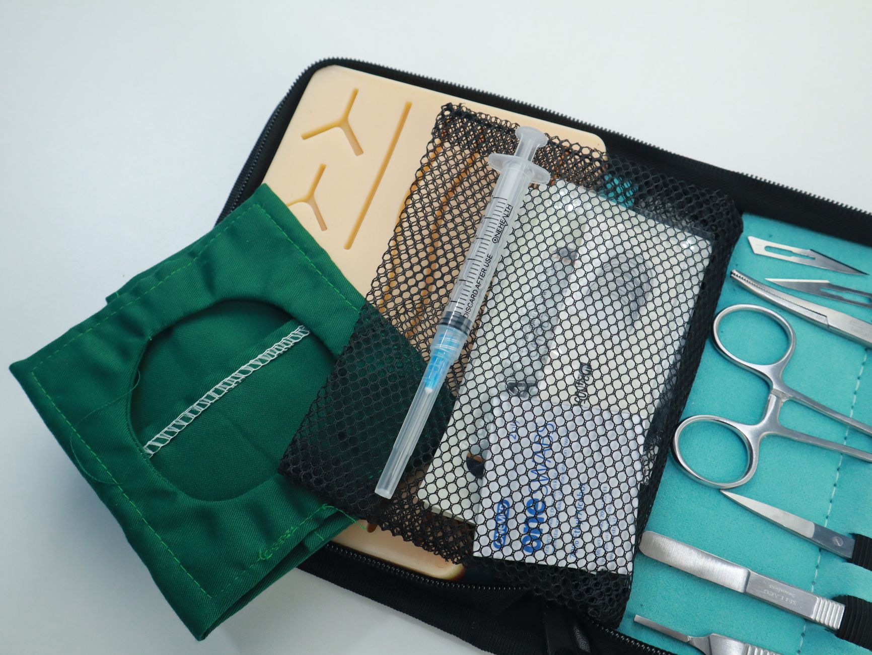
Explore how surgical simulation enhances training for anatomical variations, improving surgeon adaptability and patient outcomes.
# Training Surgeons for Anatomical Variations With Simulation
Surgical proficiency demands not only mastery of standard anatomy but also the ability to adapt to anatomical variations. These variations can significantly impact surgical approaches, complicate procedures, and influence patient outcomes. Simulation-based training has emerged as an essential tool in preparing surgeons to recognize and manage these differences effectively.
Understanding Anatomical Variations
Anatomical variations refer to deviations from the typical anatomical structures found in the general population. These can include differences in vessel location, organ shape, nerve pathways, or tissue density. While often benign, such variations can pose challenges during surgery, increasing the risk of complications if unanticipated.
### Common Examples of Anatomical Variations
- Vascular anomalies: Aberrant arteries or veins, such as a replaced right hepatic artery.
- Nerve variations: Variations in the course of the recurrent laryngeal nerve.
- Organ morphology: Differences in liver lobation or kidney position.
- Musculoskeletal differences: Variations in muscle attachments or bone structure.
Recognizing these variations preoperatively through imaging is ideal, but intraoperative surprises are common, underscoring the need for adaptable surgical skills.
Challenges in Training for Anatomical Variations
Traditional surgical training often relies on cadaveric dissection and supervised operative experience. However, cadaver specimens typically represent average anatomy and lack the dynamic aspects of living tissue. Additionally, exposure to rare anatomical variations during residency can be limited due to case variability.
These limitations can lead to:
- Reduced preparedness for unexpected anatomy.
- Increased operative time and risk.
- Hesitancy or errors in decision-making during surgery.
Role of Simulation in Addressing Anatomical Variations
Simulation offers a controlled, reproducible environment to practice surgical techniques and decision-making without patient risk. Modern surgical simulators incorporate high-fidelity models that can replicate a wide range of anatomical variations.
### Benefits of Simulation Training
- Exposure to rare variations: Simulators can be programmed with diverse anatomical scenarios.
- Safe learning environment: Trainees can make mistakes and learn from them without harm.
- Repetition and mastery: Procedures can be practiced multiple times to build confidence.
- Immediate feedback: Performance metrics and expert guidance enhance learning.
Types of Surgical Simulators for Anatomical Variations
### 1. Virtual Reality (VR) Simulators
VR simulators provide immersive 3D environments where anatomical structures can be manipulated. They allow dynamic visualization of variations and interactive procedural practice.
### 2. Physical Bench Models
These are tangible models designed to mimic specific anatomical variations. They provide tactile feedback and can be used for hands-on skill development.
### 3. Hybrid Simulators
Combining physical models with digital augmentation, hybrid simulators offer realistic tissue interaction alongside visual overlays highlighting variations.
Implementing Simulation in Surgical Training Curricula
To maximize the benefits of simulation for anatomical variations, programs should:
- Integrate variation-focused modules: Include cases with common and rare anatomical differences.
- Assess competency: Use simulation performance to evaluate readiness for complex cases.
- Encourage deliberate practice: Promote repeated sessions targeting specific variations.
- Facilitate multidisciplinary learning: Involve radiologists and anatomists to deepen understanding.
Case Study: Simulation in Laparoscopic Cholecystectomy Training
Laparoscopic cholecystectomy is a procedure where anatomical variations, such as aberrant cystic ducts or arteries, can lead to bile duct injury if unrecognized. Simulation modules presenting these variations help trainees:
- Identify variant anatomy through simulated imaging.
- Practice safe dissection techniques.
- Develop decision-making skills for intraoperative challenges.
Studies demonstrate that simulation-trained surgeons have reduced complication rates and improved operative efficiency.
Future Directions
Advancements in imaging, 3D printing, and computational modeling will further enhance simulation realism. Personalized simulation based on patient-specific anatomy may become standard, allowing surgeons to rehearse complex cases preoperatively.
Conclusion
Training surgeons to manage anatomical variations is critical for safe, effective surgical care. Simulation-based education provides an invaluable platform to develop these skills, ensuring surgeons are better prepared for the complexities of real-world anatomy.
---
Key Takeaways:
- Anatomical variations are common and can complicate surgeries.
- Traditional training may not provide sufficient exposure to these variations.
- Simulation offers safe, repeatable practice with diverse anatomical scenarios.
- Incorporating simulation into curricula enhances surgeon adaptability and patient safety.
By embracing simulation technology, surgical education can evolve to meet the demands of increasingly complex anatomical challenges.